Structural and Surface Morphological Characterization of Pure and Sr-doped TiO2 Thin Film Prepared by Spin Coating Technique
Abstract
Transparent thin films of pure TiO2 and 3% Sr-doped TiO2 (Ti0.97 Sr0.03O2) were prepared by spin coating technique onto well-cleaned glass substrate. These films were annealed at different temperatures. The structural analysis by GIXRD and Raman Spectroscopy confirms the anatase phase of TiO2. The study also shows dependence of structural parameters and crystallinity on the annealing temperature of films. Surface morphology of the prepared films studied using Atomic Force Microscopy (AFM) exhibits a homogeneous globular structure.
Keywords: TiO2 Film, GIXDR, RAMAN, AFM
Introduction
TiO2 possesses three polymorphs: anatase, rutile and brookite, with distinct crystalline structures. The rutile is the most common and well known structure of the three. In rutile, the structure is based on octahedrons of TiO2, which share two edges of the octahedron with other octahedrons and from chains Titanium dioxide (TiO2) has attracted the attention of many research workers due to its outstanding physical and chemical properties. It is found that TiO2 is also antibacterial, self cleaning and super hydrophilic. It has large number of applications as a catalyst support, gas sensor, thermoelectric and photovoltaic cells [1-13]. TiO2 is also a promising material for next generation of ultra-thin capacitors, due to its dielectric property [14-15].
With increase in the frequency, the dielectric constant of the film is found to be decrease. This is because the frequency increases dipoles start to lag behind the field and dielectric properties is going to decrease and at higher frequency dipoles are not able to follow the field for longer. The AC conductivity is linearly increase with respect to frequency and Sr-doped TiO2 show the low dielectric property and high AC conductivity compare with pure TiO2 films. An electrical property of the films has been reported [16].
Experimental & Characterization Details
In the present work, six films (three for pure TiO2; three for Sr-doped TiO2) of approximately 70-80 nm thickness were prepared. Titanium (IV) oxyacetylacetonate (0.3M) was used for Ti source and Strontium Chloride (GR) for Sr. Precursor samples were taken in polyethylene glycol (6ml) and water (4 ml). The solution was stirred for one hour at room temperature to obtain a viscous and transparent solution. The rpm of the spin coater was set at 2000 and during each rotation, three drops of solution were made to drop on the glass substrate. This process was repeated three times with the films being annealed at 3000C between each process. One film each of TiO2 and Sr-doped TiO2 were then annealed for 30 minutes at 5000C and 6000C, respectively.
The films structure was studied by Grazing Incidence X-ray Diffraction (GIXRD) using Bruker D8 discover diffractometer,(Cu kα radiation, λ=1.5406 Å) complementary information concerning film microstructure was derived from Raman Spectra which were acquired by means of a Jobin Yvon Horibra Labram- HR visible spectrometer using the blue line (488 nm) of an Ar laser as excitation source. The collection time for each spectrum was 5 minutes in the range of 100 to 200 cm-1.Film surface morphology were evaluated from AFM measurements.
Result and Discussion
Structural Characterization
GIXRD Analysis
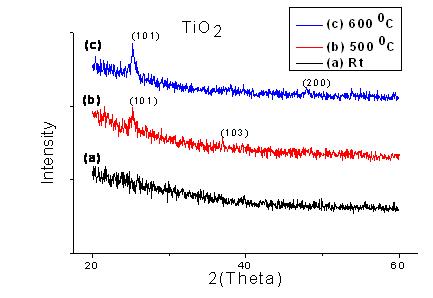
Figure 1(a), GIXRD data of the TiO2 thin films prepared by spin coating.
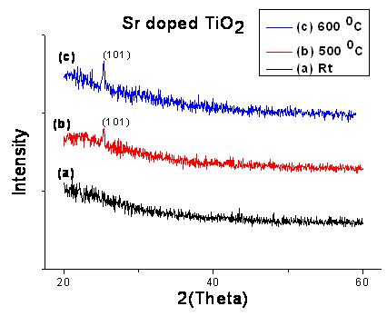
Figure 1(b), GIXRD data of the Sr-doped TiO2 thin films prepared by spin coating.
Separate peaks for Sr-doped TiO2 are not observed due to low dopant concentration. It is also difficult to predict whether the Sr ion exists as Sr-O on the surface of TiO2 as SrTiO3 from the GIXRD pattern due to low dopant concentration.
From the figure 1(a) and 1(b) it is clear that, without annealed film shows the amorphous nature and as we increase the annealing temperature the crystallinity of the film also increase. The anatase phase of TiO2 was found in both samples annealed at high temperature. The crystalline size estimate using the Debye-Scherer formula is found to be 32 nm for pure TiO2 and 37 nm for Sr-doped TiO2.
We have used short and long time scan because of very low thickness of film and we took scan in the range of 100 to 200 cm-1. The main peak of anatase phase of TiO2 is observed near about 144 cm-1. The Raman spectroscopy of the samples confirms the anatase phase of TiO2 as determined by GIXRD analysis.
Raman spectra show broadening of the spectra with increase in annealing temperature. Post annealed film also show the shifting. Same result were observed in Sr-doped TiO2 films but in whole sample we get peak near about 149 cm-1 [17-19] and they are also shows the same type of shifting compare with TiO2 Raman spectra. This result supports that the structural parameter is dependent on the annealing temperature and anatase phase is more temperature sensitive and TiO2 shows the anatase to rutile phase transformation at high temperature. The Raman shifting of Sr-doped TiO2 spectra also indicates that Sr is properly combined/attached with Ti atom.
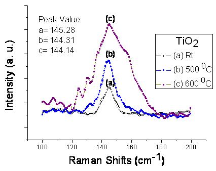
Figure 2(a), Raman spectra of TiO2 thin film prepared by spin coating technique.
The formation of TiO2 and thin films deposited by spin coating is considered accumulating like “tiny island”. Figure 3 shows the surface topography image of sample obtained by AFM. All the as-deposited thin films indicate surfaces which are not highly rough, and that the film is homogeneously distributed on the substrate. Without post annealed films show rough surface compare to annealed films. The 2D image of the films show the spherical shape of the particles and post annealed films also show same result but compare to without annealed film the particle size is small and of uniform spherical shape. The particle size calculated from the AFM data is near about ~115 nm in post annealed films in both samples.
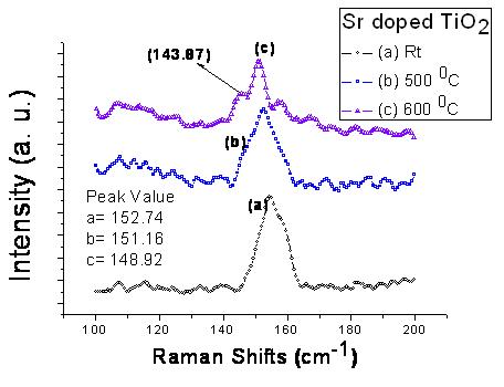
Figure 2(b), Raman spectra of Sr-doped TiO2 thin film prepared by spin coating technique.
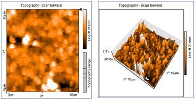
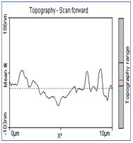
Figure 3(a), AFM data of TiO2 thin film annealed at 6000C
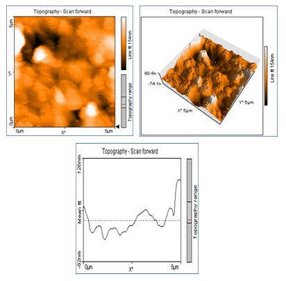
Figure 3(b), AFM data of TiO2 thin film annealed at 5000C

Figure 3(c), AFM data of Sr doped TiO2 thin film annealed at 6000C
From the AFM result, it is believed that the thin film grow process consist of three aspects, the atoms adsorption, migration and desorption, which are all connected with the annealed temperature and viscosity of the sample solutions. AFM Images obtained on different regions of the samples showed that the films exhibit a homogeneous globular structure. The entire film surface is formed by small grains of the deposited material.
Conclusion
Thin films shows the amorphous nature at room temperature (without annealed film) and post annealed film shows that the crystallinity of the films increases for the anatase phase of TiO2. Raman data suggests that the anatase phase is present in the prepared TiO2 film. The Raman spectrum shows shifting with increase in doping concentration. Raman data strongly supports the GIXRD data.Acknowledgement
Rajiv Gandhi UGC fellowship to one of the authors (M. H. Mangrola) is fully acknowledged. The authors also wish to thank UGC DAE CSR Indore, Dr. B. S. Chakrabarty (Applied Physics Department, M. S. University of Baroda), Dr. Vandana N Rao (Metallurgy & Material Science Department, M. S. University of Baroda), Dr. Utpal Joshi (School of Science, Department of Physics, Gujarat University) and Dr. M. Roy (M. L. Sukhadiya University, Udaipur) for their support and valuable suggestions.
REFERENCES :
***************************************************
M. H. Mangrola
V. G. Joshi
Department of Science & Humanity, Faculty of Engineering Technology and Research,
Department of Physics, Veer Narmad South Gujarat University,
Surat


Home |
Archive |
Advisory Committee |
Contact us